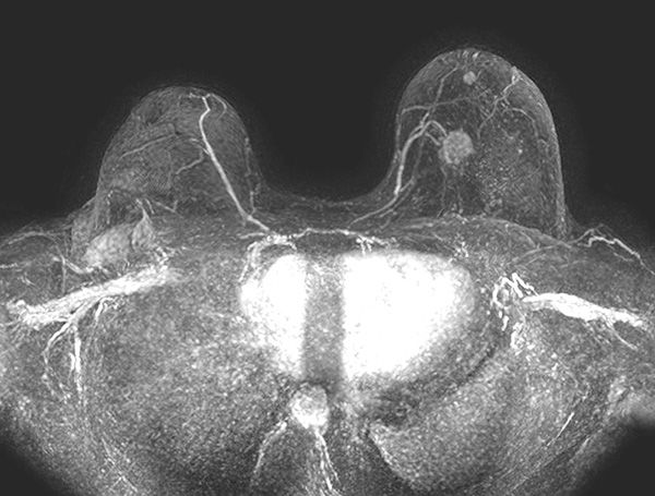
Its role is essential in the therapeutic planning of breast cancer -making it possible to diagnose non-visible lesions with other techniques – and in the follow-up of treated patients to assess recurrences. It is very useful for monitoring asymptomatic patients with a family history of breast cancer, in high-risk patients with BRCA 1 and 2 mutation, and in patients with breast prostheses.
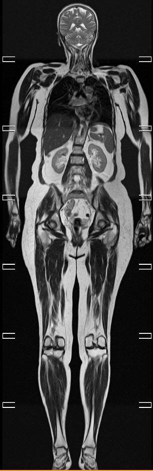
In addition to the use of MRI for the study of the breast, it is used for the study of the whole body and of the other anatomical regions: brain, spine, liver, female pelvis, etc.
Carrying out a “Total-RM” or “Whole Body Scan” makes it possible to carry out extension studies of the whole body for the detection of metastases, especially in bone. The application of new MRI sequences (diffusion, perfusion …) make MRI a more precise technique and in constant progress.